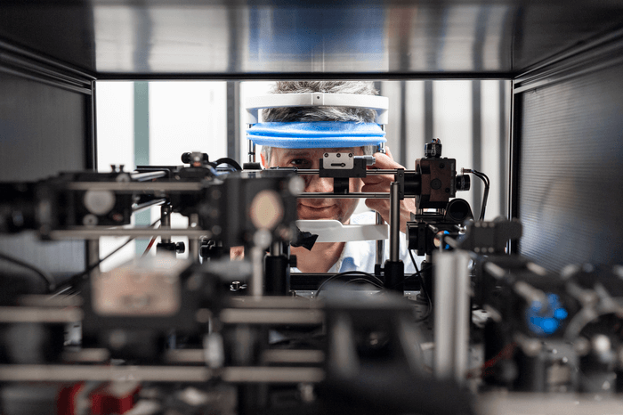
The human eye is one of the most intricate and crucial organs in the human body. Given the exceedingly fragile nature of the eye, the specific biochemical reactions that enable sight have remained a mystery. Until now, that is, thanks to a new imaging device designed by a team of scientists from the International Centre for Translational Eye Research (ICTER).
This newly created device, a two-photon excited fluorescence scanning laser ophthalmoscope (TPEF-SLO), enables the observation of molecular reactions within the eye of a living organism for the first time. This new technology could provide insights into the specific causes of degenerative eye diseases, potentially leading to new treatments and techniques for those disorders.
Capable of detecting roughly 5 million different color types, the eyes allow us to accurately assess and interact with the world around us. The molecular reactions that generate visual images occur in the retina of the eye, where photosensitive cells known as rods and cones, detect lights and colors. Rods and cones contain visual pigments, which are essential to the optimal functionality of the eye.
While the optic nerve transmits the visual signal from the eye to the brain, the biochemical process of generating visual impressions is conducted entirely within the retina itself.
“The human eye is a biochemical factory whose activity depends on biochemical transformations of a single molecule, retinal. This molecule is indispensable for the function of the visual pigments, namely rhodopsin in rods,” says Prof. Maciej Wojtkowski, who led the research, in a statement.
Since the process of converting visual stimuli, such as light and color, into visual images occurs almost solely in the eye itself, the specific molecular reactions responsible for this process have been undetectable with previous technology. Because of the delicate tissues of the eye, finding a non-invasive method to observe the biochemical reactions as they occur has proved challenging.
While traditional imaging methods have provided key insights into the changes caused by eye diseases over time, the specific mechanisms that lead to visual pigment degeneration have remained elusive.
The TPEF-SLO enables the non-invasive observation of biochemical reactions within the eye that have been invisible with previous imaging methods. This new device uses two-photon fluorescence, which allows the measurement of chemical compounds otherwise unseen by traditional single-photon fluorescence.
Metabolites of vitamin A, a crucial precursor of retinal, can be viewed with the TPEF-SLO, allowing the detection of retinal degeneration far earlier than previously possible. This, in turn, will provide countless new avenues for possible therapeutic treatments and techniques for degenerative eye diseases.
The images obtained with the TPEF-SLO have already proven effective in observing the molecular functions and changes within the eye. Through collaborations with biochemist Prof. Kris Palczewski from the University of California Irvine and the laser group of Prof. Grzegorz Sobon from the Wroclaw University of Science and Technology, the team is hopeful this new technology can be quickly applied in clinical practice.
This study was first published in The Journal of Clinical Investigation.


-392x250.jpg)


