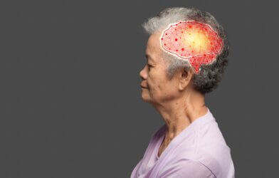
Clusters in the part of the brain called the superior colliculus demonstrate significant change depending on whether a mouse is awake or under anesthesia, according to researchers at Kyoto University. The observation of these dynamic changes suggests that the brain may optimize visual information processing depending on state of consciousness.
These clusters, which are the brain’s way of capturing visual information and converting it to an image, are often found in the visual cortex and the superior colliculus. Despite being crucial for saccades, fast eye movement between two fixed sites, and facial recognition, the evolutionarily older superior colliculus is often less studied and considered less important for visual perception.
For efficient information processing, visual information is encoded as a map-like spatial response pattern. These patterns respond to the direction and orientation of the visual image. Currently, these spatial pattern maps are not well understood, as researchers have reached conflicting conclusions in their studies.
“We revealed that these map- or cluster-like representations of moving directions were enhanced in isoflurane anesthesia,” says Masatoshi Kasai, one of the authors of the study, in a statement.
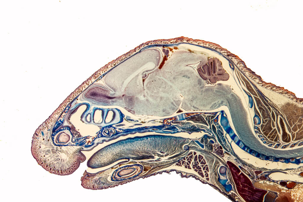
Kasai and co-author Tadashi Isa suspected that differing brain states must be the cause. To test their hypothesis, they used two-photon microscope calcium imaging to model awake and sedated states in the mice. While under anesthesia, the neurons responded at much greater volume to specific orientations as compared to the mice left awake. This result demonstrated an unexpected mapping dynamic which allows for adaptation in the superior colliculus.
“Usually, functional maps in the cortex are stable once they are formed,” says Kasai. However, Kasai adds that the results suggest “the clustering of neurons in the superior colliculus changes in response to the condition of the brain to process visual information efficiently.”
The anesthetic isoflurane was selected because, unlike other varieties, it suppresses GABAergic inhibitory neurons. According to Kasai, the pattern mapping dynamics altered by isoflurane, which have not been found with other anesthetics, further demonstrate that
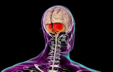
“The superior colliculus is more involved in vision information processing than previously thought,” he notes.
Kasai and his team’s findings have illuminated the significance of the superior colliculus in visual information processing. With this new understanding, future research may further explore the brain mechanisms involved in the process of understanding visual stimuli at various levels of consciousness.
The paper is published in The Journal of Neuroscience.
Article by Anna Landry


-392x250.jpg)