What if humans could camouflage their skin like a chameleon or change colors on command like an octopus? As far fetched as it sounds, understanding how these abilities originate in simple animal brains could uncover hidden talents within our own infinitely complex neural circuits.
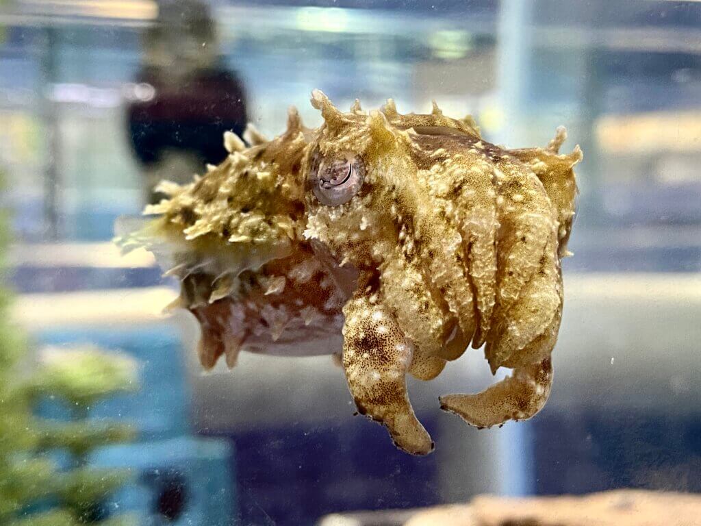
With skin that can flash like a Times Square display, the aptly named “camouflaging dwarf cuttlefish” has neuroscientists captivated. Over the past three years, researchers at Columbia University have compiled the first-ever brain atlas of this color-changing cephalopod.
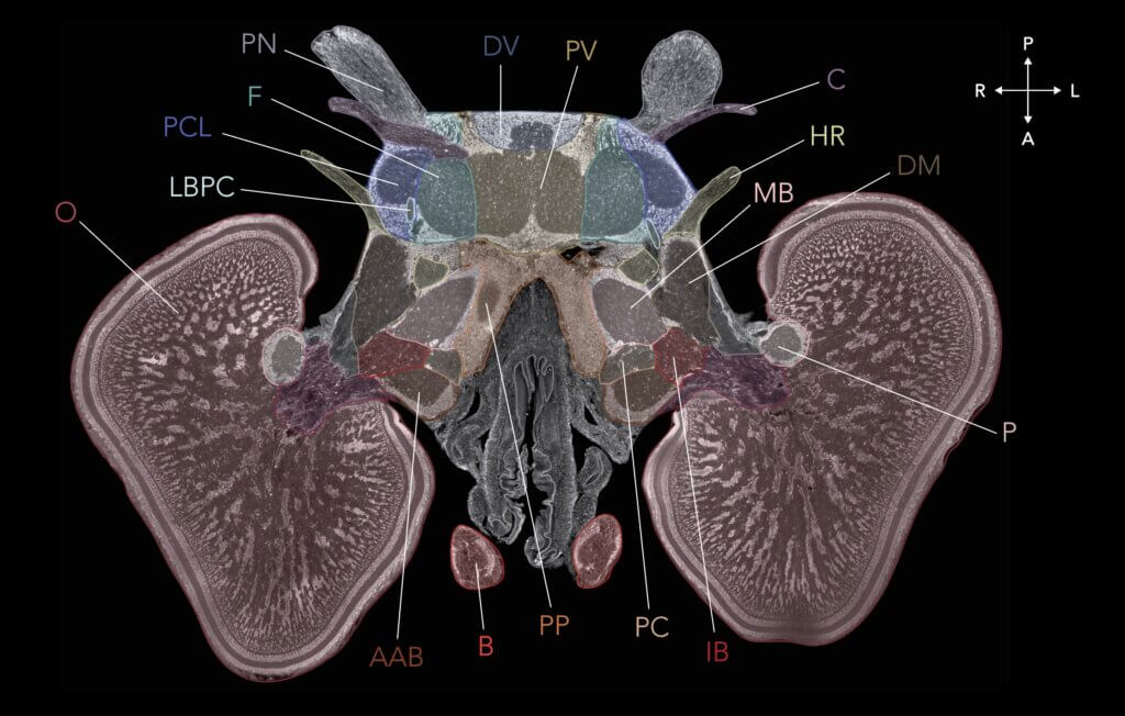
Published in the journal Current Biology, this neuroanatomical roadmap reveals for the first time the overall structure of the cuttlefish’s 32-lobed brain as well as the organization of its cells.
“One of my favorite approaches for learning about the brain is to study creatures that are highly specialized in particular behaviors, like this cuttlefish and its incredible camouflage ability,” said lead researcher Tessa Montague, PhD, a postdoctoral fellow in neuroscientist Richard Axel’s lab at Columbia, in a statement.
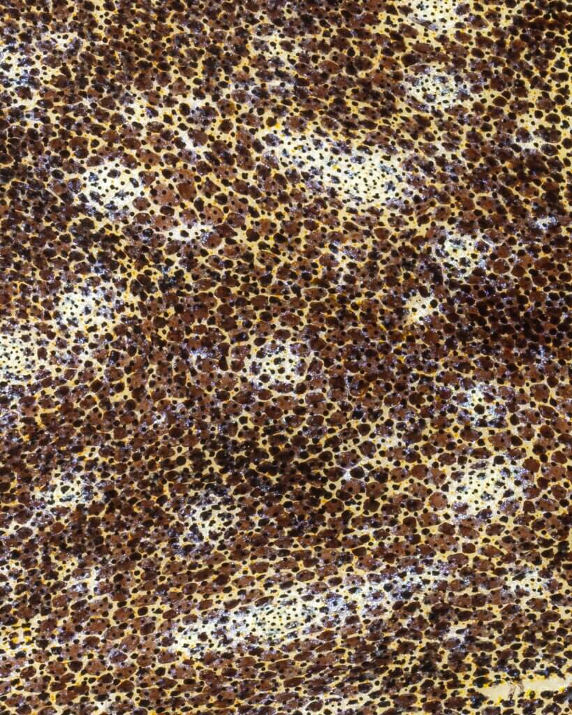
In the blink of an eye, the dwarf cuttlefish can completely transform its skin pattern and texture to disappear against its surroundings. This rapid adaptability comes from specialized cells called chromatophores that are controlled by the brain. When the cuttlefish camouflages itself, it is essentially recreating an image of its surroundings on its skin.
Study authors wanted to understand how the cuttlefish brain transforms visual input into neural signals that are then recreated as an analog image on the skin. Learning how any brain represents information and translates it into behavior is a fundamental question in neuroscience.
To do this, the researchers need to record the activity of camouflage-related neurons. But first they needed a map of the brain itself – which did not exist until now.

The Brain Atlas: A Collaborative Achievement
Constructing this intricate map required experts from several fields working together, including neuroscientists, computer programmers, web designers and anatomical imaging specialists.
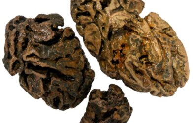
They began by using MRI scans to image the bodies and brains of eight cuttlefish. Then, using artificial intelligence, they taught a computer program to recognize the brain within the surrounding tissues in each scan. This allowed them to delineate all 32 lobes of the cuttlefish brain for the first time.
“We had developed a deep-learning approach that was able to separate the brain-related data in each MRI scan from the data linked to other tissue types in these scans. We were surprised how well we were able to adapt the technique,” said team member Sabrina Gjerswold-Selleck, who recently earned her Master’s in neurotechnology from Columbia.
Next, by matching their scans to archival microscope images of cuttlefish brains from the 1960s, the researchers painstakingly mapped out the boundaries between lobes – devoting hundreds of hours to annotate all eight brains.
This initial atlas showed the overall geography but lacked fine detail. To achieve cellular-level resolution, the team turned to staining techniques that label microscopic brain structures with different colors.
“We had a lot of back and forth about how to translate everything we had into a web-based experience that would be appealing to both scientists and nonscientists,” said neurotechnology designer Sukanya Aneja. The result is the website Cuttlebase.org, which brings the team’s data to vivid life.
A Mesmerizing Brain Revealed
With ease, Cuttlebase users can examine the intricate structure of the cuttlefish brain:
- Histological images reveal colorful neurons, glial cells and nerve fibers.
- 3D models clearly depict each brain lobe and the animal’s complex peripheral nervous system.
One can now see how the cuttlefish’s huge optic lobes process visual information from its wondrous eyes. There are also specialized lobes for motor control, memory and learning.
Even the researchers cannot resist their allure. “The cuttlefish are mesmerizing to watch,” said Dr. Montague. “When they’re camouflaging or communicating with each other, they’re effectively revealing to you on their skin what they see and how they feel.”
By creating this fascinating resource, scientists hope to reveal secrets about how all brains – even ours – represent and process information.


-392x250.jpg)