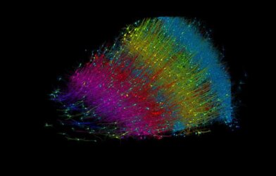
An interdisciplinary team of researchers at the University of Wisconsin-Madison has achieved a scientific first: successfully 3D-printing living brain tissue that grows, functions, and communicates like normal neuronal tissue. This breakthrough could revolutionize the study and treatment of countless neurological conditions.
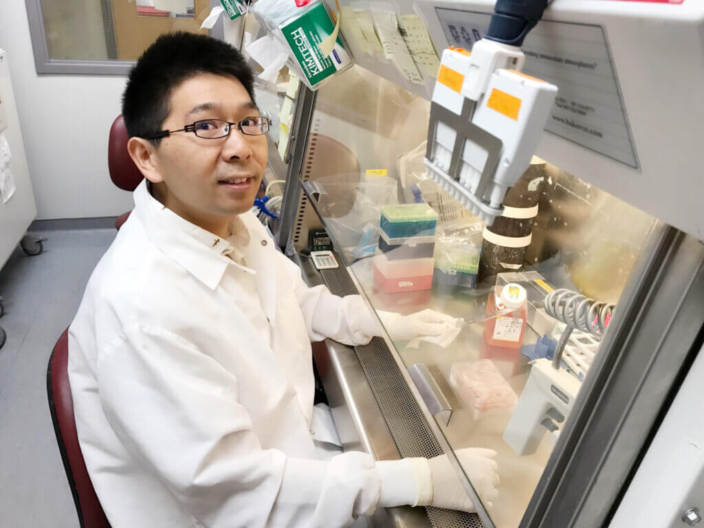
“This could be a hugely powerful model to help us understand how brain cells and parts of the brain communicate in humans,” explains Su-Chun Zhang, a neuroscience and neurology professor leading the project, in a statement. “It could change the way we look at stem cell biology, neuroscience, and the pathogenesis of many neurological and psychiatric disorders.”
So how did they do it? The key was going horizontal instead of vertical. Typical 3D-printing builds objects in stacks, layer-by-layer. But neurons don’t function that way. Zhang’s team created a special printable “bio-ink” filled with brain cells and printed it so the neurons sat side-by-side, like pencils on a table.
This configuration let the cells grow together naturally, extending tendrils to connect with neighboring cells. And connect they did—the team observed neurons communicating via neurotransmitters and forming proper networks with supporting cells, just like in the human brain.
“We printed the cerebral cortex and the striatum and what we found was quite striking,” remarks Zhang. “Even when we printed different cells belonging to different parts of the brain, they were still able to talk to each other in a very special and specific way.”
Unlike today’s most advanced mini-brains, called organoids, this printed tissue offers unprecedented cellular control and precision while remaining soft enough for oxygen and nutrients to penetrate effectively.
“Our tissue stays relatively thin and this makes it easy for the neurons to get enough oxygen and enough nutrients from the growth media,” explains Yanwei Yan, a scientist on Zhang’s team. “The tissue still has enough structure to hold together but it is soft enough to allow the neurons to grow into each other and start talking to each other.”
Such life-like function enables revolutionary applications for studying brain development, disorders, pharmacology, and more—all within a simplified and defined system.
“We can look very specifically at how the nerve cells talk to each other under certain conditions because we can print exactly what we want,” Zhang notes. “Our brain tissue could be used to study almost every major aspect of what many people at the Waisman Center are working on. It can be used to look at the molecular mechanisms underlying brain development, human development, developmental disabilities, neurodegenerative disorders, and more.”
With accessibility in mind, the group utilized common lab equipment like microscopes and electrodes to examine their creation—specialized bio-printing tools are not required. Still, they aim to refine their methods for printing specialized structures and cellular orientations.
“Right now, our printer is a benchtop commercialized one. We can make some specialized improvements to help us print specific types of brain tissue on-demand,” Yan shares.
The research is published in the journal Cell Stem Cell.