
In the quest to treat spinal cord injuries and restore function to paralyzed limbs, researchers have developed a novel approach that could revolutionize the field. A team of University of Cambridge scientists has designed a thin, flexible electronic device that can be wrapped around the spinal cord, allowing for unprecedented recording and stimulation of neural activity. This technology, known as the i360, opens up new possibilities for bypassing damaged areas of the spinal cord and restoring motor function.
The landmark findings have been published in the journal Science Advances.
The spinal cord is a crucial component of the nervous system, acting as a highway for transmitting signals between the brain and the rest of the body. When the spinal cord is injured, this communication is disrupted, often leading to paralysis below the level of the injury. Current treatments for spinal cord injuries are limited, but recent advances in neurotechnology have shown promise in restoring some degree of function.
Enter the i360, a device that takes a unique approach to interfacing with the spinal cord. Unlike traditional spinal cord stimulators, which are bulky and can only target specific areas, the i360 is thin, flexible, and designed to wrap around the entire circumference of the spinal cord. This allows for a more comprehensive and precise interaction with the neural circuitry.
“The spinal cord is like a highway, carrying information in the form of nerve impulses to and from the brain,” says co-lead study author George Malliaras, professor in the Department of Engineering at the University of Cambridge. “Damage to the spinal cord causes that traffic to be interrupted, resulting in profound disability, including irreversible loss of sensory and motor functions.”
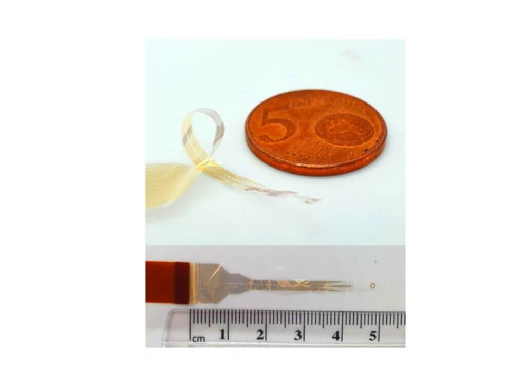
The i360 is made of a thin plastic called parylene-C, with embedded gold and titanium electrodes. These electrodes are coated with a conductive polymer called PEDOT:PSS, which enhances their ability to record and stimulate neural activity. The device is incredibly thin, measuring just 4 micrometers (about one-tenth the width of a human hair), making it highly flexible and minimally invasive.
To implant the i360, Cambridge researchers developed a surgical technique that involves carefully threading the device around the spinal cord through a small incision. This procedure was first tested in rat models, where the team demonstrated that the device could be implanted without causing damage to the delicate neural tissue. They also showed that the i360 could effectively record neural signals from the sensory and motor tracts of the spinal cord, providing a wealth of information about the activity of these pathways.
But the i360 isn’t just a passive recorder — it can also stimulate the spinal cord to elicit specific movements. By precisely targeting different regions of the spinal cord, researchers were able to selectively activate muscles in the rats’ legs, causing them to flex or extend. This level of control is a crucial step towards restoring functional movement in paralyzed individuals.
“Most technologies for monitoring or stimulating the spinal cord only interact with motor neurons along the back, or dorsal, part of the spinal cord,” explains co-lead study author Dr. Damiano Barone, from the University of Cambridge’s Department of Clinical Neurosciences. “These approaches can only reach between 20 and 30 percent of the spine, so you’re getting an incomplete picture.”
The team demonstrated that the i360 could be used to bypass a complete spinal cord injury. In a proof-of-concept experiment, they implanted two i360 devices in a rat — one above and one below a surgically induced spinal cord lesion. By recording signals from the device above the lesion and using them to trigger stimulation in the device below, they were able to restore hindlimb movement in the rat, effectively bridging the gap in the spinal cord.
While these results are preliminary and have only been demonstrated in animal models, they hold immense promise for the future of spinal cord injury treatment. The researchers are now working on scaling up the technology for use in humans, and have already tested the surgical implantation technique in human cadavers with encouraging results.
“If someone has a spinal injury, their brain is fine, but it’s the connection that’s been interrupted,” says Dr. Barone. “As a surgeon, you want to go where the problem is, so adding brain surgery on top of spinal surgery just increases the risk to the patient. We can collect all the information we need from the spinal cord in a far less invasive way, so this would be a much safer approach for treating spinal injuries.”
If successful, the i360 could offer a new lease on life for individuals with paralysis. By providing a way to bypass damaged areas of the spinal cord and restore functional movement, this technology could greatly improve quality of life and independence for millions of people worldwide.
“It’s been almost impossible to study the whole of the spinal cord directly in a human, because it’s so delicate and complex,” concludes Dr. Barone. “Monitoring during surgery will help us to understand the spinal cord better without damaging it, which in turn will help us develop better therapies for conditions like chronic pain, hypertension or inflammation. This approach shows enormous potential for helping patients.”