Thanks to CNN (no, not the cable network), scientists have been able to map the early genetic development of the human brain during pregnancy. In a study published in the journal Nature, researchers from Karolinska Institutet in Sweden are providing an unprecedented look at the genetic and molecular mechanisms that drive brain development during the first trimester of pregnancy.
Using state-of-the-art techniques, including single-cell sequencing and chromatin accessibility profiling, researchers were able to examine the brains of human embryos from six to 13 weeks after conception. This critical period is when the brain begins to take shape, with different regions forming and various cell types emerging.
“This is the first comprehensive study of brain development with a focus on gene regulation. Previous studies have almost always focused on the cortex, or cerebral cortex. Our study is a systematic mapping of the entire brain so that all regions can be compared with each other,” says lead study researcher Sten Linnarsson, professor of molecular systems biology at the Department of Medical Biochemistry and Biophysics at Karolinska Institutet.
The study focused on a process called chromatin accessibility, which refers to how “open” or “closed” certain regions of DNA are. Open regions are more accessible to the cellular machinery that reads genes, while closed regions are less active. By mapping these accessibility patterns across the entire developing brain, researchers were able to identify key genetic switches that turn on and off at specific times and locations.
Discovering the Building Blocks of the Brain
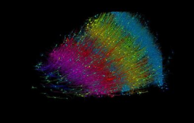
One of the most exciting findings of the study was the identification of 135 distinct cell clusters, each representing a unique cell type or state. These included various types of neurons, as well as supporting cells like glia. By comparing the genetic profiles of these clusters, the researchers were able to trace the developmental trajectories that give rise to the brain’s incredible diversity.
For example, they discovered that a group of cells called Purkinje neurons, which play a crucial role in motor coordination and balance, follow a specific developmental path. This path is guided by a carefully orchestrated sequence of gene activation and silencing, driven by special proteins called transcription factors.
“It is this process, how, in which order and in which cell types genes are activated during this process of brain formation that we have been studying. We wanted to follow the process from DNA to RNA, to the protein at each step,” explains Linnarsson.
Decoding the Language of Gene Regulation
To better understand how these transcription factors work, Swedish researchers employed a sophisticated artificial intelligence tool called a convolutional neural network (CNN). This allowed them to identify the specific DNA sequences that these proteins bind to, acting like molecular switches to turn genes on or off.
Using the CNN, they were able to decode the regulatory language that controls the development of Purkinje neurons. They found that a transcription factor called ESRRB plays a central role, activated by a two-step process involving other factors like TFAP2B and LHX5. This intricate dance of gene regulation ensures that Purkinje neurons develop properly and take their place in the complex circuitry of the cerebellum.
Beyond providing fundamental insights into brain development, the study also has important implications for understanding neurodevelopmental disorders. Many of these conditions, such as autism and schizophrenia, are thought to have their roots in the early stages of brain formation.
By linking genetic variants associated with these disorders to specific cell types and developmental stages, the researchers were able to identify potential mechanisms of disease. For example, they found that a type of neuron called midbrain-derived GABAergic neurons may be particularly vulnerable to mutations linked to major depressive disorder.
“We are now studying the onset of brain cancer in children. Fortunately, it is a rare disease, but of the various diseases that lead to death in children, it is one of the more common. We are studying the tumors that arise during embryonic brain development and using the atlas to try to understand the mechanisms of normal development that have gone wrong and how this drives tumor formation and tumor growth,” notes Linnarsson.
By providing a comprehensive map of the genetic and epigenetic landscape of the first-trimester brain, it lays the foundation for future research into the mechanisms that shape this most complex of organs.


