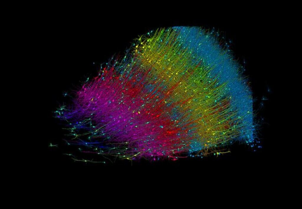
In a pioneering collaboration, researchers from Harvard University and Google have created the most detailed 3D reconstruction of a human brain fragment to date. The team has mapped a cubic millimeter of brain tissue from the human temporal cortex, revealing intricate neural connections and structures.
The achievement, recently published in the journal Science, is a significant step forward in the field of connectomics, which aims to create comprehensive maps of brain structure and connectivity.
While a cubic millimeter may seem small, the amount of data contained within is staggering.
“The word ‘fragment’ is ironic,” says study co-first author Jeff Lichtman, the Jeremy R. Knowles Professor of Molecular and Cellular Biology and newly appointed dean of science at Harvard. “A terabyte is, for most people, gigantic, yet a fragment of a human brain – just a miniscule, teeny-weeny little bit of human brain – is still thousands of terabytes.”
The mapped fragment contains approximately 57,000 cells, 230 millimeters of blood vessels, and 150 million synapses, amounting to 1,400 terabytes of data.
The team’s ultimate goal, supported by the National Institutes of Health BRAIN Initiative, is to create a high-resolution map of an entire mouse brain’s neural wiring, which would require about 1,000 times the amount of data generated from the human cortex fragment.
Google’s advanced artificial intelligence algorithms played a crucial role in reconstructing and mapping the brain tissue in three dimensions. The collaboration has also developed a set of publicly available tools that researchers can use to examine and annotate the connectome, making the results accessible to the broader scientific community.
“Given the enormous investment put into this project, it was important to present the results in a way that anybody else can now go and benefit from them,” notes Viren Jain, a Google Research collaborator.
The 3D map has revealed previously unseen details of brain structure, including rare but powerful axon connections and unusual formations such as extensive whorls. As the tissue sample was obtained from a patient with epilepsy, researchers are unsure whether these oddities are pathological or simply uncommon.
This breakthrough in brain mapping has the potential to shed light on brain function and disease, areas in which scientific understanding is still limited. The team’s next focus will be on mapping the mouse hippocampal formation, a region crucial to memory and neurological disorders.