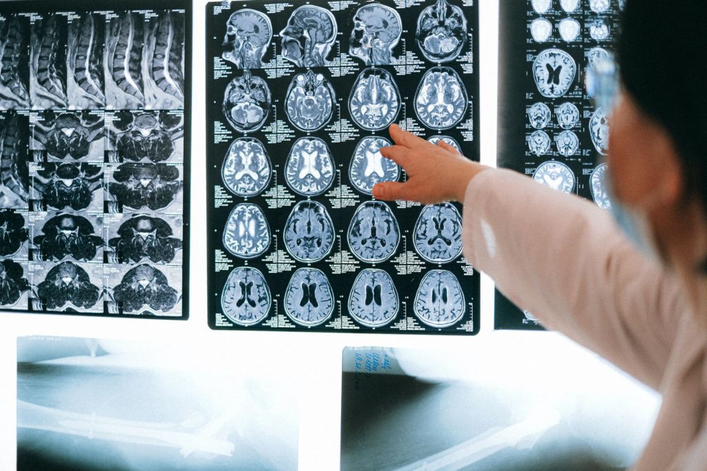
A new machine-learning tool can accurately predict the likely “brain age” of a person during an MRI brain scan, based on the amount of brain volume they have lost, according to researchers from the School of Biomedical Engineering & Imaging Sciences at King’s College London.
This new tool will allow doctors to quickly identify older-appearing brains that have lost a disproportionate amount of volume due to disease, potentially leading to earlier diagnoses and new treatment options for those afflicted, according to the study.
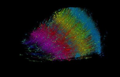
Using a convolutional neural network—a common type of artificial neural network used in deep learning that requires very little pre-processing to operate—researchers were able to accurately predict, in real-time, the likely chronological age of individuals through MRI brain scans.
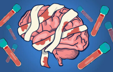
While the loss of brain volume over time is a natural part of the aging process, certain diseases, such as Alzheimer’s, will cause the brain to atrophy at a much more rapid pace. Recognizing this accelerated atrophy and determining if a patient has lost an appropriate amount of brain volume for their age, is crucial to the diagnosis of certain diseases.
But previous technology has made the real-time diagnosis of such ailments challenging, if not outright impossible. The problem is often compounded by the lack of qualified neuroradiologists at many medical centers where MRI examinations are conducted, which can create a gap between the MRI scan itself and learning the results.
“Currently, there is no screening tool during routine hospital MRI examinations that automatically detects older-appearing brains in real-time. Instead, this is assessed after the scan in centers where there are neuroradiologists, and assessment is performed visually,” said principal investigator Dr. Tom Booth in a statement.
Reducing the often inevitable delay between the MRI and analysis was a goal for researchers, who sought a real-time brain-age tool for even the most routine MRI examinations.
Researchers generated a large dataset for this study, utilizing 23,302 previous MRI brain scan examinations for patients deemed ‘radiologically normal for age.’ After accurately calculating the brain-age from this dataset, researchers applied the same brain-age prediction tool to 228 MRI brain scans that had atrophy ‘excessive for age.’ As expected, the predicted brain-age was far higher than the actual chronological age in these scans.
The use of such a broad, clinically-representative dataset was crucial to the study’s findings. “This enabled our brain-age model to generalize to out-of-sample data, like scans from external hospitals and different scanner vendors, suggesting that it can be applied in radiology centers beyond those considered in this study, particularly smaller centers without neuroradiology specialists,” said first author Dr. David Wood.
The ability to instantly determine whether an MRI brain scan reveals an excessively atrophied brain could have far-reaching implications, particularly in the study and treatment of dementia-causing diseases.
The development of this new brain-age tool will also help enhance the optimization of MRI examinations, according to researchers.
This study is published in the journal Neuroimage.
Article by Adam Swierk; Photo by Anna Shvets from Pexels


Comments