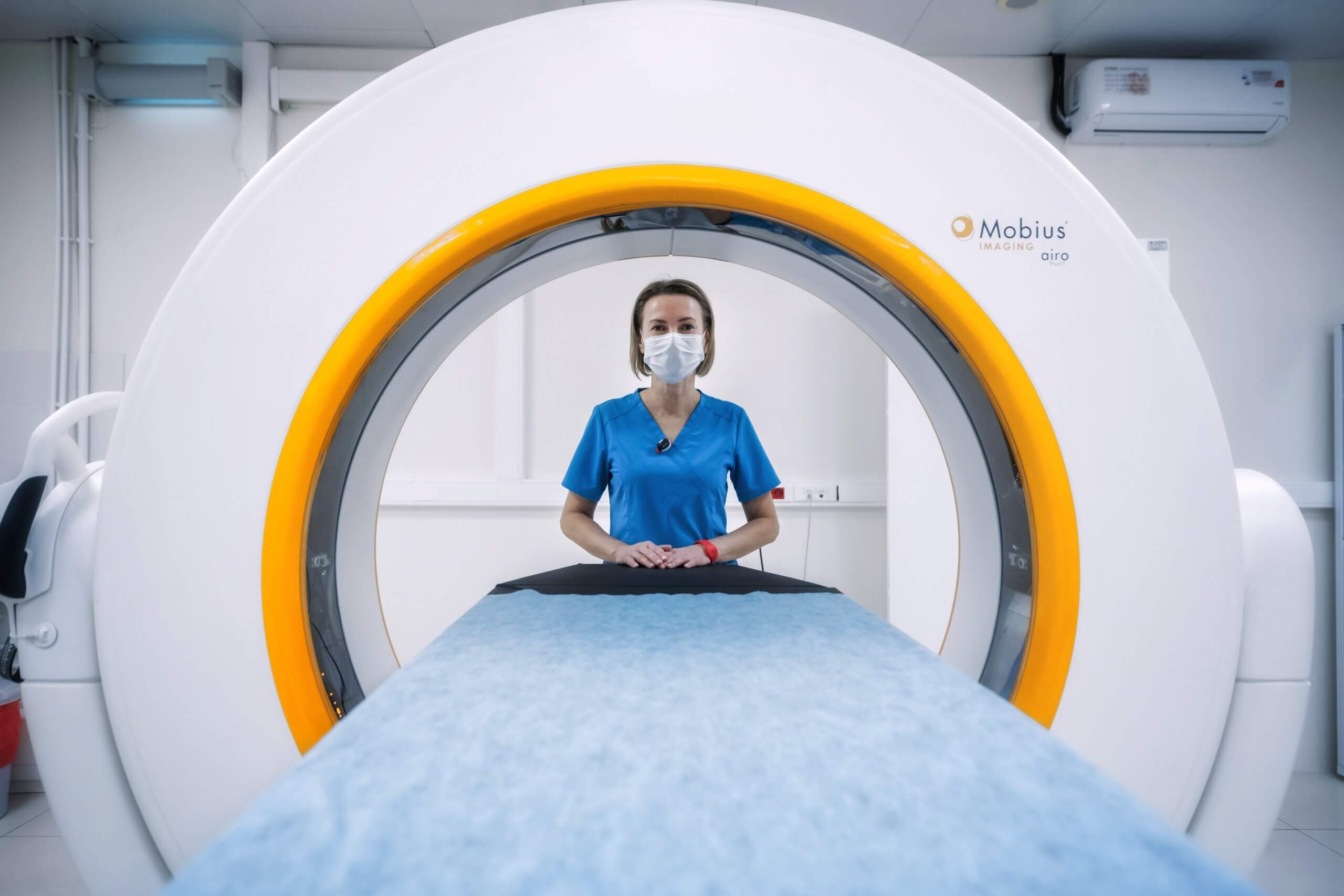
Researchers at the University of Tsukuba in Japan have made a significant breakthrough in studying human embryos using magnetic resonance imaging (MRI). They have developed a high-resolution MRI microscope that can observe human embryos at an incredibly detailed level, with a spatial resolution of one-hundredth of a millimeter.
In the world of science, understanding the development of human embryos is crucial. It allows scientists and doctors to gain insights into the growth of organs and how they function, helping to develop treatments and advance medical knowledge. Until now, conventional MR microscopes could only provide a maximum resolution of approximately four-hundredths of a millimeter, which sometimes resulted in blurry or incomplete images of tiny structures within embryos.
To overcome this limitation, the research team created an advanced MR microscope. They achieved this remarkable leap in resolution through a combination of hardware improvements, innovative imaging techniques, and efficient data collection and reconstruction methods, including the use of compressed sensing technology.
In magnetic resonance imaging (MRI), there isn’t always a direct match between the set pixel size and the actual resolution of the images. However, after rigorous testing using precisely designed artificial structures for image quality evaluation, the research team confirmed that the achieved resolution matched the set pixel size. To further validate the results, they compared the MRI image with an optical microscope image of a human embryo at the same developmental stage and found that the microstructure was accurately captured.
This groundbreaking achievement has far-reaching implications for the field of human embryology research. The enhanced resolution of this new MRI microscope allows scientists to capture the intricate details of human embryos’ microstructures. It opens the door to creating a high-resolution atlas of the brain and other vital organs during development.
Imagine having the ability to see the tiniest structures within an embryo with unprecedented clarity. This technology promises to revolutionize our understanding of human embryology, providing researchers with the tools they need to create detailed maps of how our brains and organs form and grow.
This breakthrough isn’t just about capturing images; it’s about unlocking the secrets of life itself. It’s a testament to human ingenuity and our relentless pursuit of knowledge. With this powerful tool in their hands, scientists are poised to make discoveries that could lead to better treatments for various medical conditions and ultimately improve human health.
In the world of science, every tiny detail matters, and this high-resolution MRI microscope is a giant leap forward in our quest to unravel the mysteries of life before birth. It’s a reminder that even the smallest things can have the most significant impact on our understanding of the world around us.