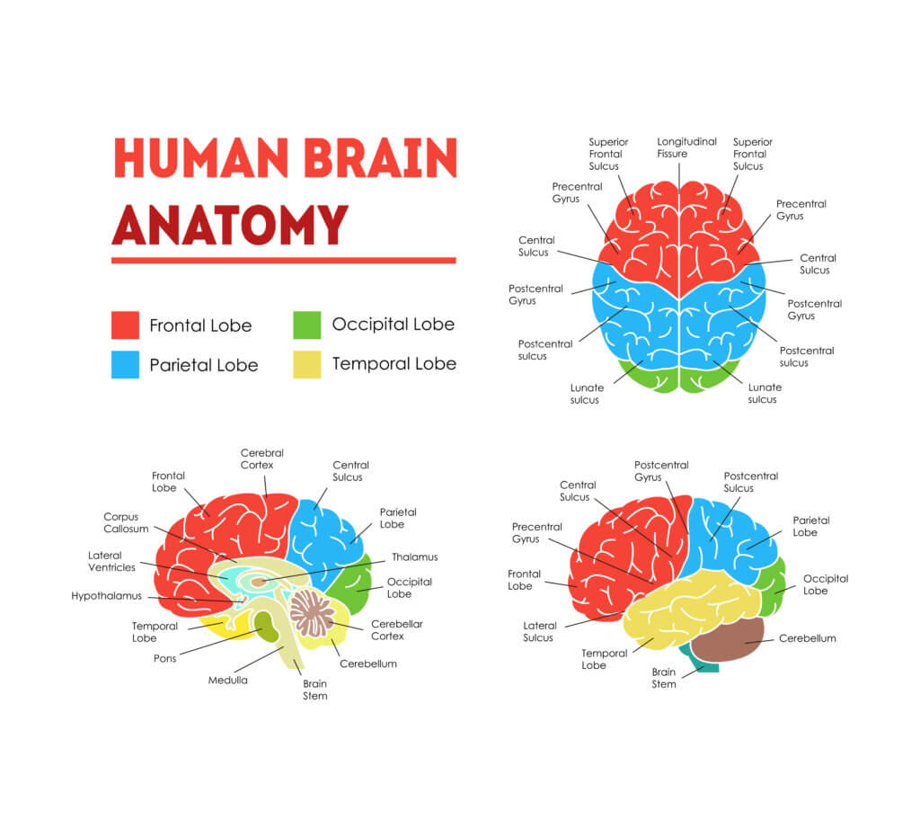
To study the anatomy of the brain, we did some dissection in cadavers, but the bulk of study was in an atlas, with the brain cut in cross section, every 2 to 3 millimeters. They showed structures within the brain with catchy names, like the marginal portion of sulcus cinguli and the flocculonodular lobe. Some of us studied together at the library, walking there, uphill, both ways, in deep snow. Year-round. Today’s young whippersnappers simply tap into their iPads. I hope you’ll take a few moments to breathe in the brain’s magnificence.
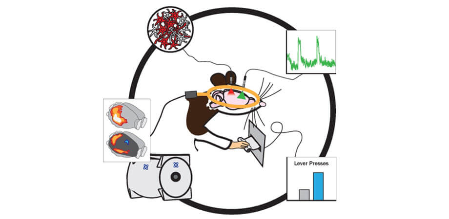
It has long been the quest of scientists to simultaneous manipulate and monitor living brain activity, to associate groups of cells with particular behaviors. It’s happening, thanks to a hybrid of optogenetics and functional magnetic resonance imaging (fMRI).
Optogenetics combines genetic and optical methods to enable study of brain function and behavior via manipulation or monitoring of specific cell groups, brain regions, or neural pathways. fMRI maps activities across the whole brain. These two technologies as “opto-fMRI” have a powerful synergy, fueling neuroscientific research.
Scientists at Pennsylvania State University recently published an overview of the history, technical advances, applications, and important considerations of optogenetics and fMRI, and their synergistic combination. The future applications of this hybrid method in neuroscience and neuroimaging are of impressive scope.
Optogenetics and fMRI have each, alone, made significant contributions to the expansion of scientific knowledge. Since 2010, advanced technology has enabled the joining of the two methods, providing valuable contributions to both neuroscience and neuroimaging. The combination expands our knowledge of brain regions from just the anatomical to the functional.
Opto-fMRI is an important tool for examining therapeutic methods and their applications, such as deep brain stimulation (DBS), to treat disorders such as Parkinson’s disease, chronic pain, and obsessive-compulsive disorder. Much more study is needed to understand the uses of opto-fMRI for studying global brain functioning.
The research is published in the open-access journal Neurophotonics.