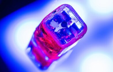
In ancient times doctors used spiderwebs to make bandages for their patients. In 2022, there is a 3D model of living brain cancer. It was developed as a potential alternative for testing drugs in development without using lab animals. In its creation, researchers at KTH Royal Institute of Technology in Stockholm, Sweden conquered one of the biggest challenges in tissue engineering.
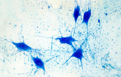
The researchers reported a technique for replicating the body’s smallest blood vessels, the microvasculature, inside of collagen hydrogel loaded with living cancer cells. The technique, called cavitation molding, creates cavities small enough for cells to form into blood vessels on a scale more closely resembling those of the human body.
Alessandro Enrico, a PhD student at KTH, says that the technique is a breakthrough in biomedical research and that the method could potentially be used for modeling other kinds of human tissues.
Previously, the only alternative to animal testing was simple 2D cell models, in which human cells are cultured on plastics in a flat, two-dimensional arrangement. Enrico says that although “lab-on-a-chip” platforms in 2D are used to replicate living tissue but are limited by their simplicity.
“The 2D tissue models slow down testing and makes it more expensive,” Enrico says in a statement. “The results of working with a 3D model relate to the actual 3D dimensional tissue in the human body.”
The 3D tissue models bridge the gap between simple 2D models and live tissue physiology, he says.
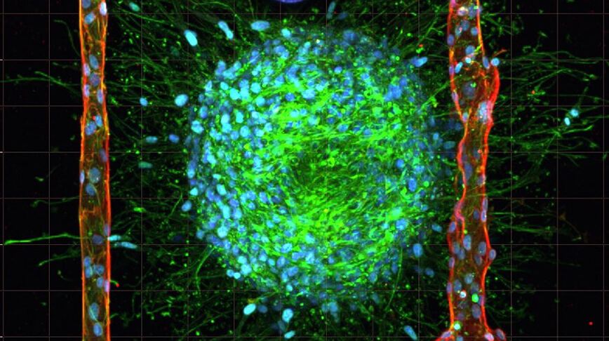
Enrico says the first step was casting an unstructured collagen hydrogel containing living cancer cells. Then, they used laser irradiation of the hydrogel to generate gas bubbles that rearrange the collagen fibers, creating cavities and shaping microchannels. Endothelial cells are pumped into the cavities, where they assemble into artificial blood vessels similar in size to the natural vasculature of the human body.
The 3D tissue models are stable for at least eight days in physiological conditions. “This is essential for studying complex biological interactions that can take days or weeks to develop,” Enrico notes.
Researchers say next step is developing models of different tissues and organs.
The findings are published in the journal Advanced Materials.