A new set of brain sensor grids are capable of reading the electric signals from the surface of the brain at a resolution 100 times higher than currently available, according to a new study.
Given the incredibly delicate nature of the human brain, surgery on the vital organ is a vastly complex endeavor. The use of electrocardiography (ECoT) sensor grids on the brain’s surface is already in widespread use by surgeons. ECoT provides crucial information in preparation for tumor removal and epilepsy surgeries.
If approved for clinical use, the new ECoT sensor grids will monumentally improve the level of electric signal details provided from the brain’s surface. This will refine the accuracy of brain surgeries in the future, ensuring healthy tissue is less likely to be affected or removed during procedures, according to the study.
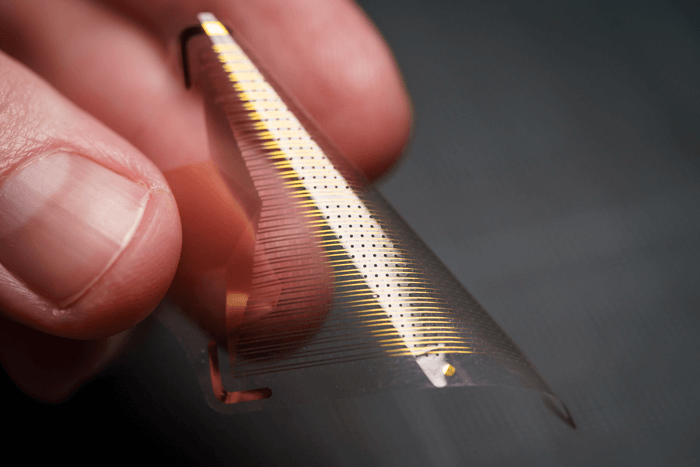
While current ECoT sensor grids only hold between 16 and 256 total sensors, the new sensor grids developed by researchers are embedded with either 1,024 or 2,048 ECoT sensors. This drastic increase in sensors per grid was made possible by the reduction of space between ECoT sensors, from one centimeter apart to one millimeter.
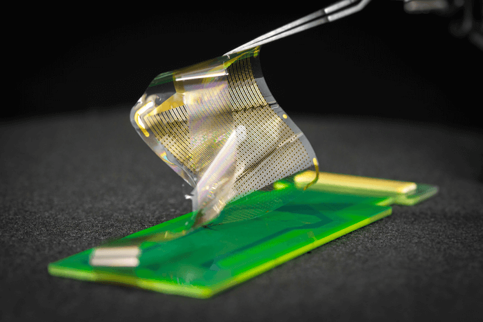
Not only do the new ECoT sensor grids provide far more details regarding electric brain signals, but they are also 100 times thinner than currently-approved sensor grids. The use of nanoscale platinum rods in the construction of the new sensor grids makes them more flexible than currently used models, allowing them to cover a wider surface area when applied to the cerebral cortex.
The unique manufacturing process for the new ECoT sensor grids permits a wide variety of customization in both shape and size, according to the study. The supremely customizable nature of the new ECoT sensor grids will allow surgical teams to create more personalized, functional maps of each patient’s brain, as well.
Additionally, since the new ECoT sensor grids were designed with clinical use in mind, customized sensor grids can be created with holes in them. This means the new ECoT sensor grids can be used during the actual surgical procedure rather than solely in advance, maximizing a surgeon’s potential for accuracy and precision.
While the new ECoT sensor grids are currently being targeted for clinical use by brain surgeons, researchers are hopeful their new technology can allow for wireless models to be developed in the future. This could have a profound impact on treatment options for those with unmanageable epilepsy and potentially lead to improvements in the quality of life for patients living with paralysis or neurodegenerative disorders.
The team also hopes their research could provide a pathway towards further insights, and increase our overall understanding of how the brain functions and operates moving forward.
Shadi Dayeh at the University of California San Diego Jacobs School of Engineering led the project in conjunction with a team of specialized doctors, engineers, and researchers.
The research is published in Science Translational Medicine.
Article by Adam Swierk